By Debbie Bunch
March 10, 2025
Lung Expansion Interventions Fall Short for Abdominal Surgery Patients
Do lung expansion interventions reduce the number of long-term respiratory complications among patients undergoing abdominal surgery?
According to a study conducted at 17 academic medical centers nationwide and led by investigators from Massachusetts General Hospital and the University of Colorado Anschutz Medical Campus, the answer is no.
Researchers randomized abdominal surgery patients to either a lung expansion set of interventions or usual care. The bundle included:
- Preoperative education on postoperative pulmonary complications (PPCs).
- Intraoperative protective ventilation with individualized PEEP to maximize respiratory system compliance.
- Intraoperative neuromuscular blockade administration and reversal based on the patient’s weight and neuromuscular transmission monitoring.
- Postoperative supervised incentive spirometry and mobilization encouragement.
All the patients in the study were at intermediate or high risk for PPCs and were assessed for a range of respiratory issues for 90 days following their surgery. The investigators tracked everything from slightly low oxygen levels to needing a breathing machine.
While patients who received the lung expansion interventions did require fewer rescue treatments for poor oxygenation during surgery, the positive impact did not extend to the follow up period, where no difference in respiratory complications was seen between the two groups. However, many of the patients did suffer from respiratory complications.
“The costs associated with treating these complications are in the order of billions of dollars,” said study author Marcos F. Vidal Melo, MD, PhD. “We’ll have to think outside the box to prevent them.” The study was funded by the National Heart, Lung and Blood Institute and published by The Lancet Respiratory Medicine. Read Press Release Read Abstract

Watching Lungs Grow in Real Time Could Lead to Better Care for Preemies
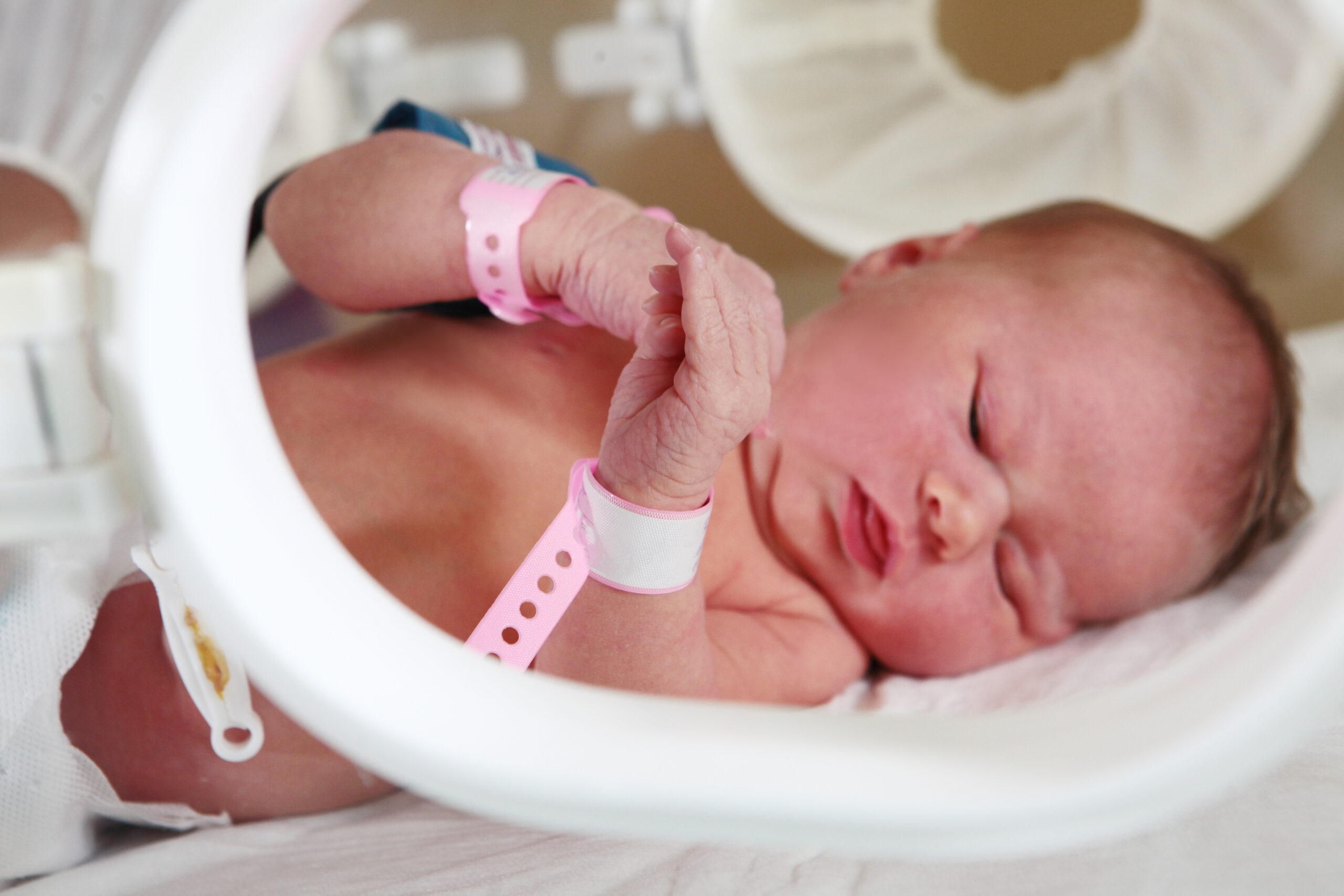
Researchers from Vanderbilt University and Vanderbilt University Medical Center believe they are on the path to understanding how injured lung tissue in premature infants may heal and regenerate — and it’s not what they thought.
The authors note respiration occurs in the lungs’ alveoli across a fragile basement membrane between epithelial cells and blood vessels. Traditional wisdom says ingrowing septa emerge from a layer of epithelial, endothelial, and mesenchymal cells to divide airspaces into alveoli.
Their study used a four-dimensional microscopy technique to create 3D video images of mouse lung tissue grown in the laboratory. When the researchers watched the mouse lung’s development unfold, they witnessed a ballooning outgrowth of epithelial cells supported by a ring of myofibroblasts—cells that promote tissue formation.
They believe this new technology will lead to the identification of specific molecules and pathways that guide this process and serve as a discovery tool for drugs that can promote tissue regeneration after injury – advances that could help the 50% of infants born two to four months prematurely who develop bronchopulmonary dysplasia.
“If we can understand how the lung forms, then we have a blueprint for how to grow new lungs after injury,” said study author Nick Negretti, PhD.
The National Institutes of Health (NIH) supported the study and published in JCI Insights.
Read Press Release Read Full Paper

Injectable Therapy for Lung Damage May Be on the Horizon
A proof-of-concept study conducted by researchers from the Perelman School of Medicine at the University of Pennsylvania might one day help heal damaged lungs. The injectable therapy, which consists of mRNA and a new lipid nanoparticle, can travel directly to the lower regions of the lung.
“The lungs are hard-to-treat organs because both permanent and temporary damage often happen in the deeper regions where medication does not easily reach,” explained study author Elena Atochina-Vasserman, MD, PhD. “Even drugs delivered intravenously are spread without specificity. That makes a targeted approach like ours especially valuable.”
Like the COVID-19 mRNA vaccines, the injectable therapy uses lipid nanoparticles as the mRNA delivery system. In this case, the investigators specifically matched mRNA with ionizable amphiphilic Janus dendrimers, lipid nanoparticles derived from natural materials found in previous studies to be organ specific, making them a good candidate to send mRNA explicitly to the lungs.
Once the mRNA is in the lung, it instructs the immune system to create transforming growth factor beta, a signaling molecule vital for the body to repair tissue.
“This research marks the birth of a new mRNA delivery platform with its strengths and potential beyond the original mRNA LNPs,” said 2023 Nobel laureate Drew Weissman, MD, PhD, a study co-author.
Dr. Weissman notes that the new platform is also easier to deal with because, unlike the COVID-19 mRNA vaccines, it does not have to be stored at extremely cold temperatures and is easier to produce.
The team is also exploring the use of the platform in the spleen.
The study was supported by the NIH’s Institute of Environmental Health Sciences and published by Nature Communications.


